Fisica a Ferrara: orientamento didattico
Medical Physics – Research Activities
Medical Physics – Research Activities
Medical Physics
Research Activities
X-ray Imaging

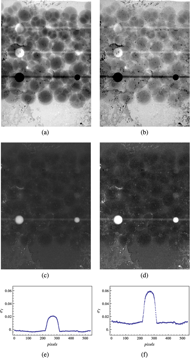
X-ray imaging is one of the most reliable among the non-invasive diagnostic techniques currently available in modern medicine. Indeed, it allows the radiologist to view the details of the internal structures of our body with a very fine spatial resolution (order of a tenth of a millimeter). Concerning Breast imaging, the most recent technological innovations now allow for the three-dimensional reconstruction of the anatomical tissue, and through the use of contrast agents it is also possible to identify tumors in the early stage of their development, thus leading to more effective treatments. Our commitment is towards the optimization of these techniques, so as to provide radiological imaging with the best tools for diagnosis and prevention. Furthermore, the medical use of ionizing radiation represents the main contribution to the radiation dose among the population of the European Union. Training of the Medical Physics Expert is one of the EU strategies to contain this risk, and our research group has been involved in the European network EUTEMPE to address this important educational task.
Innovative X-ray Sources
In the field of X-ray imaging, the radiation source plays a fundamental role. To improve image quality, while simultaneously decreasing the delivered radiation dose, it is important to develop innovative sources of monochromatic X radiation. Monochromatic sources, unlike traditional sources such as X-ray tubes, emit radiation of a single energy. Over the years the group has been involved in various projects with this goal, both national and European, such as SL-Thomson@INFN-LNF, Labsynch, Eurogammas@ELI-NP, and currently MariX. The main interest has been focused on the realization of inverse Compton sources for medical applications. Inverse Compton sources are based on the interaction between accelerated electron beams and intense laser beams. The research group deals with the study and measurement of the characteristics of the radiation beams and development of innovative techniques, in order to optimize the results of diagnostic imaging, through Monte Carlo simulations and experimental activities in the department laboratories.
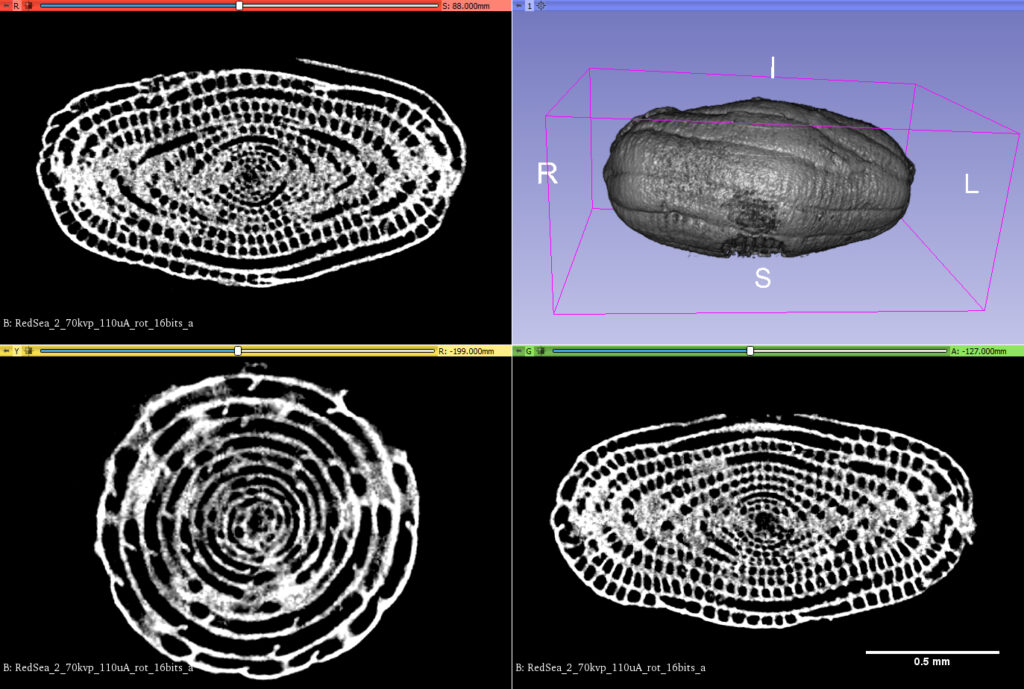
X-ray Microtomography
X-ray microtomography (XMT) is a non-destructive evaluation technique that allows the internal structure of an object to be imaged by reconstructing the spatial distribution of the local linear X-ray absorption coefficients of the materials/phases contained within. This provides a virtual 3D representation of the internal architecture of an object from which two-dimensional (2D) cross-sectional slices can be extracted along the three orthogonal planes of an object. The 3D object can be converted into a microstructurally faithful 3D mesh suitable for Finite Element Modelling that can describe the geometry of each constituent. Micro-CT has applications both in medical imaging and in industrial computed tomography. In general, there are two types of scanner setups. In one setup, the X-ray source and detector are typically stationary during the scan while the sample/animal rotates. The second setup, much more like a clinical CT scanner, is gantry based where the animal/specimen is stationary in space while the X-ray tube and detector rotate around. These scanners are typically used for small animals (in vivo scanners), biomedical samples, foods, microfossils, and other studies for which minute detail is desired.
Nuclear Medicine
The widespread availability of novel radioactive isotopes showing nuclear characteristics suitable for diagnostic and therapeutic applications in nuclear medicine (NM) has experienced a great development in the last years, particularly as a result of key advancements of cyclotron-based radioisotope production technologies. At Legnaro National Laboratories of the National Institute of Nuclear Physics (LNL-INFN), Italy, a 70-MeV high current cyclotron has been recently installed. This cyclotron will be dedicated not only to pursuing fundamental nuclear physics studies, but also to research related to other scientific fields with an emphasis on medical applications. LARAMED project was established a few years ago at LNL-INFN as a new research line aimed at exploiting the scientific power of nuclear physics for developing innovative applications to medicine. The goal of this program is to elect LNL as a worldwide recognized hub for the development of production methods of novel medical radionuclides, still unavailable for the scientific and clinical community. Although the research facility is yet to become fully operative, the LARAMED team has already started working on the cyclotron production of conventional medical radionuclides, such as Tc-99m, and on emerging radionuclides of high potential medical interest, such as Cu-67, Sc-47, and Mn-52.
Radionuclides Radiotherapy
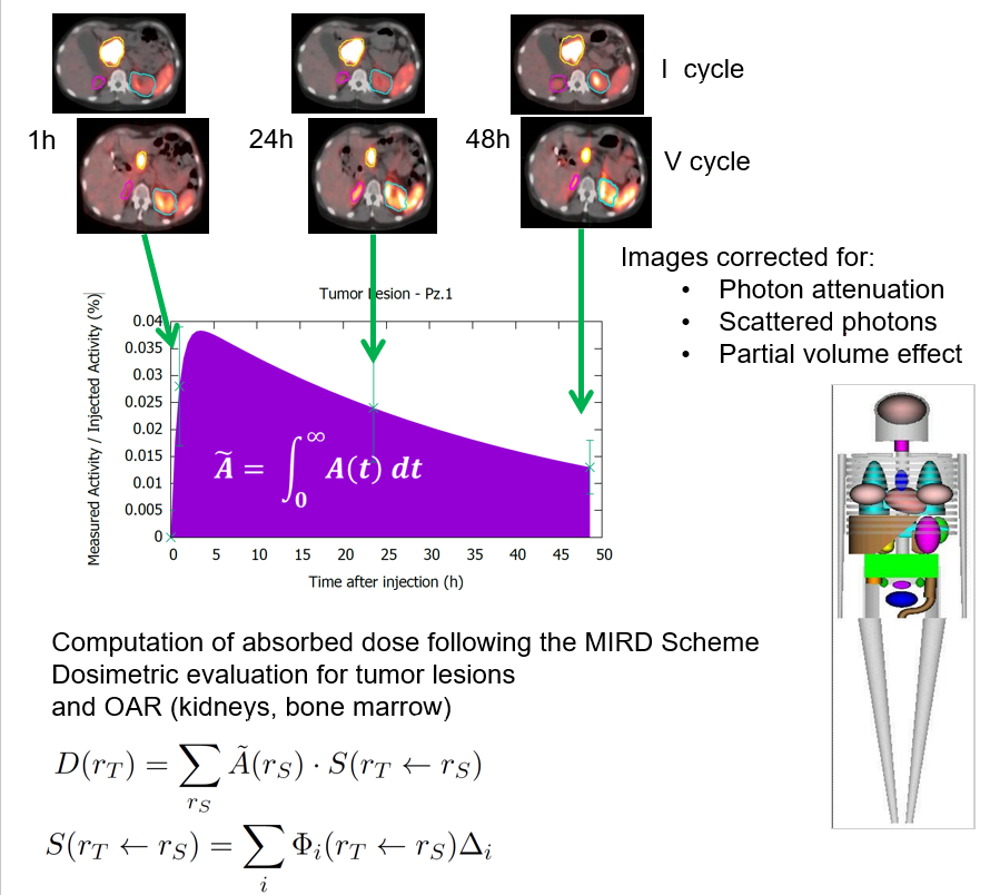
Targeted radionuclide therapy (TRT) is a promising technique for cancer therapy. The therapeutic agent is administered to the patient via different methods: intravenous injection, oral uptake or direct intra-tumoral injection. The aim of TRT is to deliver a lethal dose of radiation to tumours from internal sources by targeting their specific molecular or functional targets with agents. Compared to a conventional chemotherapy or external radiotherapy, it has the potential to deliver therapeutic radiation more specifically to cancerous in a way that minimizes toxicity to surrounding normal tissues. Advanced imaging techniques, especially radionuclide imaging like SPECT or PET, can be used to determine the spatial distribution of administered tracers for calculating the organ-absorbed dose. During the last years there has been a growth of TRT due to several new isotopes and radiopharmaceuticals for the treatment of metastatic bone pain, neuroendocrine and other solid tumours, mainly in the liver. Therefore, clinical practitioners would like to have a priori-knowledge of the biodistribution and washout of TRT agents before TRT. This information enables optimization of the therapeutic efficacy by adjusting the administered dose to each patient and can be obtained non-invasively by single or sequential nuclear medicine scans, e.g., planar and emission computed tomography (ECT) or combined with blood sampling if necessary. Quantitative images at different time points allow fitting of time activity curves for the targeted organs and tumours. From the integration of these time-activity curves one can obtain the cumulative activity and further convert it to the absorbed dose, which can help pre-treatment planning or post-therapy dose verification.
Modelling and Monte Carlo simulations of particle interactions with matter
Monte Carlo (MC) simulations play an important role in a variety of situations in medical physics. In particular, MC codes that simulate the interactions of particles with matter can be exploited to predict the outcome of an experiment or to refine the design of a component of the experimental setup. The effect of changing a system parameter can be studied without the need of a real irradiation of the sample/patient, which can be prevented due to dosimetric issues or simply because the radiation source under investigation is not yet available. The Medical physics group is involved in various Monte Carlo studies, spanning from those related to new source of radiation, such as inverse Compton sources, to those that aim to model the response of a detector. The studies are carried out through Geant4, which is a C++-based open-source particle tracking code, among the more complete and powerful. A project the group is involved is AGATA, which is funded by INFN and aims at developing a framework for performing virtual clinical trials in breast x-ray imaging by means of digital models of patients developed from high-resolution 3D breast images and a MC simulation software. The developed platform will permit to evaluate the performance of various imaging apparatus and modalities without the need of irradiating the patient.
In order to increase the field of application and make the simulations closer to reality, the physical models have to be refined and extended. An important research area of the group is the development of new models of the coherent interactions of X-rays with matter and their implementation in Geant4. Among the considered phenomena there are the inclusion of interference effects in Rayleigh scattering of X-rays in both amorphous and crystalline materials and the X-ray refraction/reflection at the interface between two different materials. These extensions open the way to inclusion of phase effects in Geant4, which can then be used to simulate phase-contrast imaging. The extension of Rayleigh scattering model be found at the link: www.fe.infn.it/medical_physics/Geant4/Rayleigh_MI.
Study of Cerebral Venous Return
The aim of this research topic is to characterize the human cardiovascular function by developing advanced non-invasive protocols easily usable in clinic and research, in co-operation with the Vascular Diseases Centre of the University of Ferrara. Such activity is based on the fruitful results of the Drain Brain project, successfully conducted by our group aboard the International Space Station, where the circulatory system functionality of a crewmember was assessed. In the framework of the new project named WISE – Wearable non-Invasive SEnsors for cardiovascular function assessment, we are involved in the development of an open ultrasound and plethysmography system to obtain simultaneous traces with high flexibility to be integrated with current standardized measurements (e.g. electrocardiography). Moreover, we are developing mathematical models for the simulation of human blood flows and pressures. The cited devices and techniques will be put in action together for in-vitro measurements by using ad-hoc developed phantoms, in cooperation with the CRIBO Training Academy.
Laboratories and Research Facilities
The work of the Medical Physics research group includes several experimental activities, which are carried out at the laboratories available at the Physics and Earth Science Dept. of Ferrara University:
LARIX
Large Italian X-ray facility, which hosts the X-ray equipment including several X-ray experimental lines equipped for the test of innovative X-ray imaging techniques, experimental activity for didactics on X-ray imaging, monochromatization by Bragg diffraction, as well as a microfocus X-ray tube dedicated to micro-tomography research.
Nuclear Medicine Laboratory
The Nuclear Medicine Laboratory contributes to the design, development and construction of advanced detectors for application to radioactive source detection and measurement. In the last years, the laboratory contributed to build a SPECT/CT scanner for preclinical imaging, now in use at “Dipartimento di Morfologia, chirurgia e medicina sperimentale” – UNIFE and a Beta Counter based on TDCR principle for absolute activity measurement of pure beta radionuclides.
Eco-fluid dynamics Laboratory
The Echo-fluid dynamics Laboratory hosts advanced ultrasound devices and materials for the development and test of innovative dedicated phantoms, experiments about the non-invasive assessment of the cardiovascular status in humans, as well as experimental activity for training on echo-Doppler imaging.